An outline of oogenesis and spermatogenesis
Gametogenesis is the overall term given for the production of gametes, the egg and sperm cell. Oogenesis is the process where the ovum or egg cell is made. Oogenesis is the female type of gametogenesis where the opposite is spermatogenesis for the male production of sperm cells. Mitotic division is where the cell replicates the DNA before splitting into identical daughter cells with a full set of chromosomes or diploid. All cells divide through mitosis but in animal cells, only gametes divide by a process called meiosis where the cell divides to give daughter cells which have half the number of chromosomes or haploid cells, (Brevini, 2013).
· Humans have 46 chromosomes
· Cows have 60 chromosomes
· Pigs have 38 chromosomes
· Chickens have 78 chromosomes
· Sheep have 54 chromosomes
· Horses have 64 chromosomes
(Lawrence et al, 2012).
Oogenesis starts when germ cells migrate to the ovaries and the follicular cells begin to surround the germ cells and becomes an oogonium. The oogonium goes through mitotic division and produces many oogonia. This happens in the very early stages of life, before birth. The oogonium goes through oocytogenesis which creates the primary oocyte through mitosis. The two daughter cells, the primary oocytes, have a full set of chromosomes.
Ootidogenesis is the next step where the primary oocyte divides thorough meiosis. The process of Ootidogenesis stops at prophase 1. This therefore means that the female is born with a finite number of primary follicles. Further development of the primary follicle begins of the expansion of the oocyte in the adolescent stages of life. The surrounding follicular cells begin to replicate and become many layers thicker. This is called a granulosa. The granulosa secrets certain glycol-proteins and cross link to form a zona pellucida. By the time of ovulation the oocyte goes through meiosis and generates a polar body. This body deteriorates and the egg leaves the ovary. In the oviduct the egg begins the second division begins. This halts at the metaphase stage until the egg is fertilised, (Albertini et al, 2013).
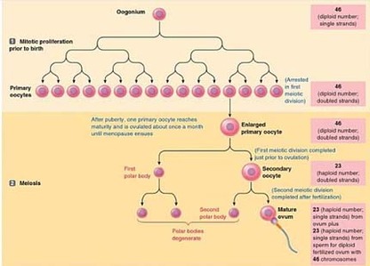
Figure 4 (Sherwood, 2010)
Spermatogenesis
There are over one billion spermatogonia in the tesicles. This forms the base layers of the germinal epithelium. There are two types of cells which can be identified. Type A cells are the spermatogonia which undergo mitotic division. This is how the spermatogonia numbers are kept at around one billion.
Spermatogenesis begins in the seminiferous tubules when the second group of type A cells go under spermatogenesis process. They are connected by thin bridges of cytoplasm and are preserved in these cytoplasmic connections. These cells go through a mitotic division and where one cell becomes a type B cell and the other daughter cell replenishes the spermatogonia. The B type cells then undergo another mitotic division and become primary spermatocytes (I).
The newly divided primary spermatocytes (I) will now enter the first stages of meiosis. In this stage they replicate the DNA. The cells then migrate towards the special milieu of the luminal compartment. They undergo a complex stage of meiosis and begin to differentiate from each other and begin to be visible in a light microscope. This process takes approximately 24 days. Following the long prophase stage, metaphase, anaphase and telophase stages are relatively shorter. Two secondary spermatocytes are created.
A second meiosis occurs and the secondary spermatocytes divide into spermatids. This can occur quickly as DNA recombination does not occur. This process lasts a few hours and forms two spermatids with only half the number of chromosomes and DNA.
The spermatids mature, with the help of Sertoli's cells are actively converted into sperm cells in the epididymis of the testies, (Frandson et al, 2009).
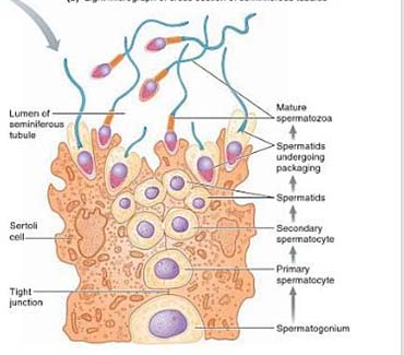
Figure 5 (Sherwood, 2010)
